
Atlas of sectional anatomy of man. Bone-muscular system. Melller T.B., Ryf E.
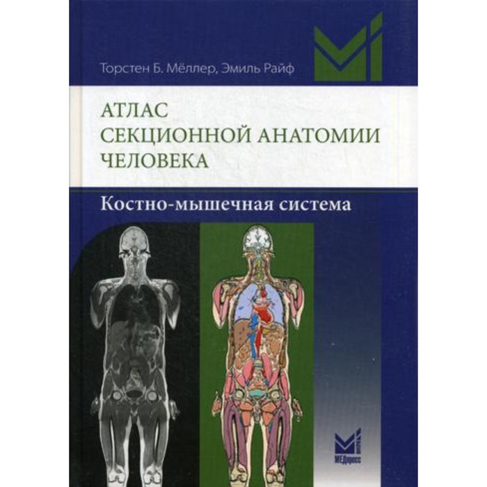
This book is a solid work created on the basis of great experience and deep knowledge of the authors in the field of MRI diagnostics, which uses the original method of presenting the material. Magnetic resonance tomograms were obtained on an MR tomograph with an induction of a magnetic field of 3 T and are distinguished by clarity and a high degree of detail. All tomograms are accompanied by semi -chimatic color illustrations made personally by the authors, with the color coding of anatomical structures, and at the bottom of each page a scheme is given that indicate the direction and side of the body. The illustrations strictly fit into a certain concept, reflect only the co-muscular structures under consideration, significantly facilitate the use of the atlas and, of course, are its dignity. The order in which the illustrations are presented allows you to easily find the desired cut.
The publication begins with the overview images of the whole body and the temporal-nominal joint obtained using 32-channel radio frequency coils. The remaining images are distributed into three chapters: about the spine, the upper limb and the lower limb. The last two chapters provide images of the cross sections of the upper and lower limb from the proximal departments to the distal, ensuring the continuity of the perception of anatomical structures. The atlas used the officially adopted terminology.
Atlas is designed for radiation diagnostics, surgeons, traumatologists, anatomes.
| Characteristics | |
| A country | Russia |
| Author | Melller Torsten B. |
| Number of pages | 292 |
| The year of publishing | 2018 |
| Type of cover | Hard cover |
| View | Specialized medicine |
There are no reviews for this product.